Arteriovenous Malformation Explained
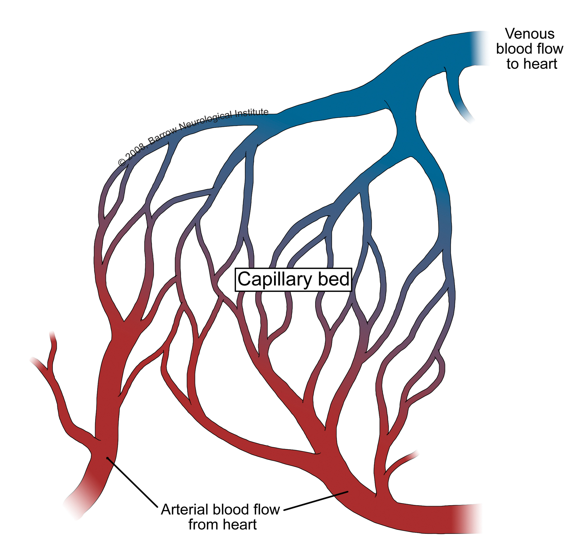
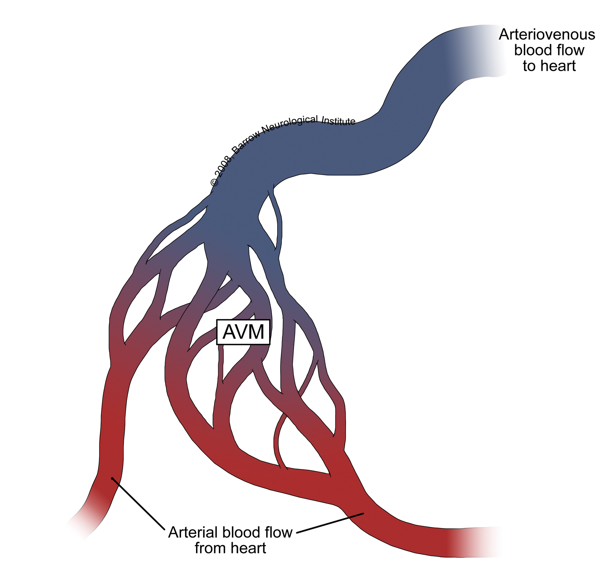
An arteriovenous malformation (AVM) is a complex tangle of abnormal arteries and veins linked by one or more direct connections called fistulas or shuts. This tangle of abnormal arteries and veins is referred to as a nidus. Normally, as the high-pressure arterial blood is pumped through a capillary bed there is a gradual decrease in blood pressure before reaching the venous system. With an AVM, the capillary bed is absent and the high-pressure arterial blood bypasses normal brain tissue and is pumped directly into the normally low-pressure venous system.
There is typically high blood flow through the nidus of the AVM, but it is not known whether the flow is a cause or effect of the abnormal blood vessels, or both. One thought is that the high-pressure blood from the arterial system gravitates towards the path of least resistance. Another thought is that the AVM itself recruits blood vessels.
Ultimately, the arterial blood rushes through the AVM, instead of working through available capillary beds, which feed the surrounding brain tissue, increasing blood flow through the nidus. This re-direction of the arterial blood away from the brain tissue and through the AVM is referred to as shunting.
Over time, the high blood flow and shunting of high-pressure arterial blood through the AVM causes the feeder arteries and veins making up the AVM to dilate or expand. This dilation weakens veins making them susceptible to hemorrhage and the arteries susceptible to aneurysms.
AVMs are thought to be congenital, arising from developmental derangements at the embryonic stage of vessel formation, at the fetal stage. However, this has never been clearly established and they may arise after birth. AVMs are usually single, except when associated with hereditary hemorrhagic telangiectasia (HHT).
- AVMs occur in <1% of the population and their cause is unknown
- AVMs are thought to be due to abnormal development of blood vessels in utero and may be present since birth
- An estimated 300,000 Americans have AVMs
- About 12% of people with an AVM experience symptoms
- Each year, 2-4% of people with an AVM have a hemorrhage
- About 2% of hemorrhagic strokes are related to an AVM
- In around 50% of people with an AVM, an intracranial hemorrhage is the first sign
Schematic representation of an arteriovenous malformation with a draining vein and a vessel that enters the AVM on the right without passing through the AVM and a vessel en passant that skirts the AVM. *Used with permission from Barrow Neurological Institute.

Normal capillary blood flow occurs when oxygenated arterial blood from the heart branches through smaller vessels and deoxygenated blood is collected in the veins and delivered back to the heart. *Used with permission from Barrow Neurological Institute

The blood flow of an AVM is abnormal; oxygenated arterial blood from the heart makes direct connections to large draining veins, no intermediate capillary bed. *Used with permission from Barrow Neurological Institute.
Symptoms May Include
- Seizures
- Headache
- Stroke-like symptoms (weakness, paralysis, numbness, tingling, vision, balance and hearing problems)
How Are They Diagnosed?
Most AVMs are detected with either a computed tomography (CT) brain scan, a magnetic resonance imaging (MRI) brain scan or a cerebral angiogram.
Treatment is offered to try to prevent bleeding from the AVM. Your doctor will recommend the best treatment for you and this will be determined by the size and the location of your AVM.
SPETZLER-MARTIN AVM Grading System

The Spetzler-Martin Grading System assigns a score of 1 for small AVMs (3 cm). The eloquence of adjacent brain is scored as either non-eloquent (0) or eloquent (1). The venous drainage is scored as superficial only (0) or including drainage to the deep cerebral veins (1). Scores for each feature are totaled to determine grade. Class A combines Grades I and II AVMs. Class B are all Grade III AVMs. Class C combines Grades IV and V AVMs.
The Spetzler Martin Grading Scale estimates the risk of open neurosurgery for a patient with AVM, by evaluating AVM size, pattern of venous drainage, and eloquence of brain location. A Grade 1 AVM would be considered as small, superficial, and located in non-eloquent brain, and low risk for surgery. Grade 4 or 5 AVM are large, deep, and adjacent to eloquent brain. Grade 6 AVM is considered not operable.
- Size of nidus
- small (<3cm) = 1
- medium (3 – 6cm) = 2
- large (> 6cm) = 3
- Eloquence of adjacent brain
- non-eloquent = 0
- eloquent = 1
- Venous drainage
- superficial only = 0
- deep = 1

